Histology Of Compact Bone Diagram : HLS [ Cartilage and Bone and Bone Histogenesis, compact ... / Describe the histology of bone tissue.. It can be remodeled all throughout life to. Bone tissue is regulated by several hormones including 3. Compare and contrast compact and spongy bone. Cartilage and bone are specialized connective tissues that provide support to other tissues and organs. Video on histology of compact bone from anatomy diagram courtesy :
Histology of compact bone is shown along with osteons, haversian canals, volkmann's canals, osteocytes, lacunae, and canaliculi. Woman koi fish tattoo rib cage. The osteon or haversian system /həˈvɜːr.ʒən/ (named for clopton havers) is the fundamental functional unit of much compact bone. The extracellular matrix consists of about 15% water, 30% collagen fibers, and 55% crystallized mineral salts like calcium. It is the shell of many bones and in the histology of normal bone, as a result of the normal remodeling process, up to 20% of the bone surface may be covered by osteoid (usually 10 µm thick).
Compact Bone Diagram . Compact Bone Diagram Spongy Bone ... from i.pinimg.com A cross section of a typical osteon or haversian system. The human eye can discern only two types of bone. Given below is a labeled diagram to help you understand the structure of compact long bones, as well as the microscopic structure or histology of the haversian system of compact bones. It is found beneath the periosteum of all bones and makes up the bulk of the diaphyses of long bones. It is the shell of many bones and in the histology of normal bone, as a result of the normal remodeling process, up to 20% of the bone surface may be covered by osteoid (usually 10 µm thick). Available at the itunes store and for android users at the google play store. Bone is never formed as a primary tissue: Compact bone, microscopically, is made of numerous osteons, whereas spongy bone is composed of sheets of lamellar bone and does not contain osteons.
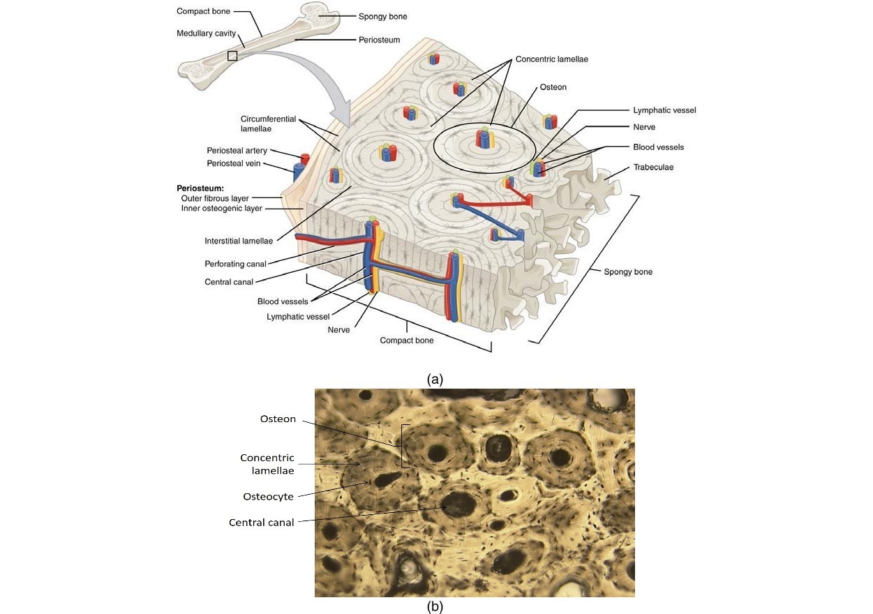
Given below is a labeled diagram to help you understand the structure of compact long bones, as well as the microscopic structure or histology of the haversian system of compact bones.
Compact bone, microscopically, is made of numerous osteons, whereas spongy bone is composed of sheets of lamellar bone and does not contain osteons. Bone tissue is regulated by several hormones including 3. Osteons are roughly cylindrical structures that are typically between 0.25 mm and 0.35 mm in diameter. These haversian systems have long cylindrical haversian. Learn vocabulary, terms and more with flashcards, games and other study tools. Compact bone and spongy/cancellous bone are the two types of bones in the human body. There are two types of bone formation: If the outer layer of a cranial bone fractures, the brain is still protected by the intact inner layer. Shotgun histology bone cells and matrix bone is a tissue in which the extracellular matrix has. Compare and contrast compact and spongy bone. In development there are 2 separate signaling pathways for pattern formation and the formation of bone itself. This ossification process can be studied in the long bones, such as the. Video on histology of compact bone from anatomy diagram courtesy :
These haversian systems have long cylindrical haversian. Skincare terbaik untuk kulit berminyak dan berjerawat. Though bone comes in several shapes and sizes the structure and composition of bone is the same in all. Aftershokz air bone conduction headphones. Osteons are roughly cylindrical structures that are typically between 0.25 mm and 0.35 mm in diameter.
5.4: Bone Structure - Medicine LibreTexts from med.libretexts.org Osteons are roughly cylindrical structures that are typically between 0.25 mm and 0.35 mm in diameter. A cross section of a typical osteon or haversian system. In development there are 2 separate signaling pathways for pattern formation and the formation of bone itself. Compact bone high resolution histology diagram. Video on histology of compact bone from anatomy diagram courtesy : Compact bone stands in stark contrast to trabecular bone in several ways. Normal bone histology & embryology 101 with dr. The differences between compact and spongy bone are best explored via their histology.
Video on histology of compact bone from anatomy diagram courtesy :
Long bone, compact bone and spongy bone. Skincare terbaik untuk kulit berminyak dan berjerawat. The functional units of compact bone are osteons; These haversian systems have long cylindrical haversian. You may also save it to your computer for more zoomed view. The human eye can discern only two types of bone. The hollow region in the diaphysis is called the medullary cavity, which is filled with yellow marrow. The histology of compact bone. The strongest form of bone tissue. Woman koi fish tattoo rib cage. It can be remodeled all throughout life to. The osteon or haversian system /həˈvɜːr.ʒən/ (named for clopton havers) is the fundamental functional unit of much compact bone. The diaphysis is the tubular shaft that runs between the proximal and distal ends of the bone.
Bone is never formed as a primary tissue: Feel free to use for study purposes. Long bone, compact bone and spongy bone. Aftershokz air bone conduction headphones. It always replaces a preexisting support tissue.
Compact Bone Diagram : The stability of a compact bone is ... from www.histology.leeds.ac.uk You may also save it to your computer for more zoomed view. Aftershokz air bone conduction headphones. Compact bone stands in stark contrast to trabecular bone in several ways. From the compact bone histology slide, i will enlist some important histological features that you might identify at the laboratory. Long bone, compact bone and spongy bone. This shows the architecture of compact bone which is designed to nourish and regulate osteocytes and bone matrix. Start studying histology of compact bone. Click on the image to enlarge it.
Cartilage and bone are specialized connective tissues that provide support to other tissues and organs.
Here we see the microscopic structure of bones that contains an extracellular matrix that surrounds cells. You may also save it to your computer for more zoomed view. The human eye can discern only two types of bone. This shows the architecture of compact bone which is designed to nourish and regulate osteocytes and bone matrix. Compact bone consists of outer and inner sheets of lamellar bone (not seen here) and haversian systems, shown here, that run parallel to the long axis of bones. Normal bone histology & embryology 101 with dr. It can be remodeled all throughout life to. Mammalian compact bone is composed mostly of haversian system. Cartilage occurs where flexibility is required, while bone resists deformation. • spongy bone lies internal to the endosteum and comprises a network of lamellae that do not form the haversian channels and osteons found in compact bone. Long bone, compact bone and spongy bone. It is called the haversian system. In the compact bones, the bone cells are arranged in a particular pattern.
Learn vocabulary, terms and more with flashcards, games and other study tools compact bone diagram. In development there are 2 separate signaling pathways for pattern formation and the formation of bone itself.


0 Komentar